



AI Advances Low-Field MRI Toward Clinical Use
1 Abstract
In recent years, with the continuous maturation of hardware design and the application of AI technology in image reconstruction and enhancement, the imaging quality and clinical application potential of low-field MRI have been steadily improved, attracting renewed attention. Compared with high-field MRI, low-field systems feature lower costs, more flexible deployment and greater accessibility. However, they also face challenges such as low signal-to-noise ratio, insufficient spatial resolution and prominent artifacts. The advancement of deep learning has provided a new technical pathway for image quality improvement of low-field MRI. This review systematically summarizes two core applications of deep learning in low-field MRI: denoising and super-resolution. It covers the technical evolution from supervised U-Net and generative adversarial networks (GANs) to diffusion models (DMs), and discusses practical challenges including data dependency and interpretability. Overall, low-field MRI is gradually evolving from a "low-cost alternative" into an imaging platform with unique clinical value, and deep learning serves as a key driving force for this transformation.
2 Denoising Models: Supervised and Unsupervised Learning
2.1 Supervised Denoising: U-Net Architecture and Its Extensions
U-Net is one of the most prevalent backbone structures for low-field MRI denoising. Its encoder–decoder design with skip connections can extract features while preserving high-frequency details to the maximum extent, making it highly suitable for medical image restoration. In related studies, 3D anisotropic U-Net has been adopted for image quality transfer (IQT) from low-field to high-field images, achieving favorable restoration performance across multiple datasets. The Denoising Autoencoder (DAE) further simplifies the architecture and is suitable for cross-field-strength transfer. To address electromagnetic interference, a common issue in portable MRI, residual U-Net has been used to directly predict interference-free signals, demonstrating strong practicality.
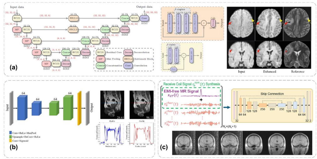
Figure 1. Architecture of anisotropic U-Net
Besides image-domain approaches, some studies perform reconstruction or denoising directly in the k-space. For instance, AUTOMAP realizes end-to-end learning of the mapping from raw signals to images and exhibits good robustness in ultra-low-field systems. Other works combine classification models, visualization methods and transfer learning to jointly optimize artifact identification, signal recovery and fast imaging. In general, supervised denoising methods have evolved from simple image restoration toward greater emphasis on structural fidelity and cross-scenario adaptability.
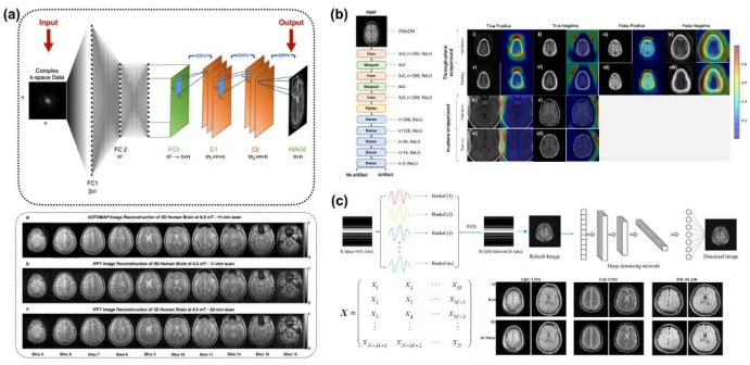
Figure 2. Network architecture of AUTOMAP
2.2 Supervised Denoising: U-Net Architecture and Its Extensions
Since supervised learning relies on paired data, which is not always readily available in clinical practice, unpaired learning circumvents this limitation. GAN and CycleGAN are representative methods, enabling image translation from low-field to high-field MRI in the absence of strictly paired datasets.

Figure 3. Workflow of Cycle-GAN for LF-MRI denoising (two generators + two discriminators)
On this basis, several studies further introduce residual blocks, structural loss and multimodal evaluation to improve anatomical consistency of reconstructed results. Diffusion models have become a hot research topic in recent years. Compared with traditional GANs, diffusion models deliver superior generation stability and detail recovery, opening new possibilities for zero-shot enhancement of low-field MRI. Nevertheless, such methods usually demand higher computational resources and involve relatively complex inference pipelines. Therefore, balancing performance and efficiency remains an important direction for future research.

Figure 4. Diffusion model
3 Deep-Learning-Based Super-Resolution Methods
3.1 Supervised Super-Resolution: CNN and Residual Learning Architectures
The core goal of super-resolution is to recover fine structural details from low-resolution low-field images. This is critical for lesion identification, tissue segmentation and quantitative analysis. In this field, U-Net, DenseNet, residual learning and attention mechanisms are widely deployed. Some studies implement image super-resolution and synthesis via cascaded 3D U-Net, and further integrate segmentation tasks to boost consistency of outputs. Other works utilize multi-channel input and multi-scale feature extraction to better recover structural details such as infant brain tissues.

Figure 5. Architecture of supervised super-resolution network
In addition, some models unfold the iterative framework of compressed sensing into learnable networks, combining data consistency with deep feature extraction to achieve a good trade-off between reconstruction speed and image quality. Other research adopts densely connected convolutional blocks and attention fusion modules to further strengthen structural recovery capability. In short, supervised super-resolution is no longer merely "image upscaling", but gradually develops toward the integration of reconstruction, enhancement and clinical segmentation.
3.2 Self-Supervised Super-Resolution: GAN Implementation
Under limited data conditions, GAN-based methods offer more flexible solutions for low-field super-resolution. The advantage of such frameworks lies in the ability to pre-train on large-scale high-field datasets first and then adapt using a small amount of low-field data, improving model usability in real low-field environments. This strategy carries strong practical significance for low-field MRI characterized by scarce data and complex scenarios.
4 Discussion
Research priorities for deep learning in low-field MRI have shifted from merely demonstrating method feasibility to investigating generalization, robustness and clinical usability in real-world settings. Data preparation remains the foundation; high-quality annotations, reasonable data augmentation, unified normalization and noise labeling strategies all directly affect model performance. In terms of model selection, U-Net remains the most mature and commonly used backbone. However, hardware conditions vary greatly among different low-field systems, so there is no universal architecture applicable to all scenarios.
Generalization is also one of the most concerning issues. Excellent performance on single-center datasets cannot be directly translated into multi-center clinical applications. Furthermore, interpretability constitutes a major barrier to clinical acceptance. As models grow increasingly complex, enabling clinicians to understand the reasoning behind model predictions has become an indispensable step for translating deep learning for low-field MRI into clinical practice. Meanwhile, the application potential of low-field systems for specific populations and scenarios deserves further exploration.
Overall, the field still faces common challenges including data scarcity, insufficient model interpretability, inconsistent evaluation criteria and limited public benchmark datasets. It is certain, however, that with the coordinated advancement of hardware and algorithms, the clinical value of low-field MRI is continuously being redefined.

Innovation Academy for Precision Measurement Science and Technology, CAS.
West No.30 Xiao Hong Shan, Wuhan 430071 China
Tel:+86-27-8719-8631 Fax:+86-27-8719-9291
Email:hanyeqing@wipm.ac.cn
鄂ICP备15017570号-1
