

75 Pioneering Stories on the Path to Technological Self-Reliance and Strength —— Lighting Up the Lungs! The Journey to Breakthroughs in High-End Domestic MRI Equipment
"We need this equipment urgently; can it be quickly installed at Jinyintan Hospital?" During the COVID-19 pandemic, the lungs were the most affected organ in infected patients. Clearly visualizing changes in lung function was crucial for understanding the virus's damage to lung physiology and advancing clinical treatments. In February 2020, at the critical moment of Wuhan's fight against the pandemic, Zhang Dingyu, then-director of Wuhan Jinyintan Hospital, learned that the Innovation Academy for Precision Measurement Science and Technology at the Chinese Academy of Sciences had developed a "human lung multi-nuclei magnetic resonance imaging system." He immediately voiced the need for the equipment. "Fully support! If one machine isn't enough, find a way to send more!" Upon receiving this urgent request from Wuhan's frontline, the Party Leadership Group of the Chinese Academy of Sciences promptly responded with firm instructions. This domestically developed high-end medical device, led by a research team from the Chinese Academy of Sciences, played a vital role on the frontline of the pandemic. However, few people know that the team behind this lung-illuminating device dedicated over a decade of rigorous research to bring it to fruition.
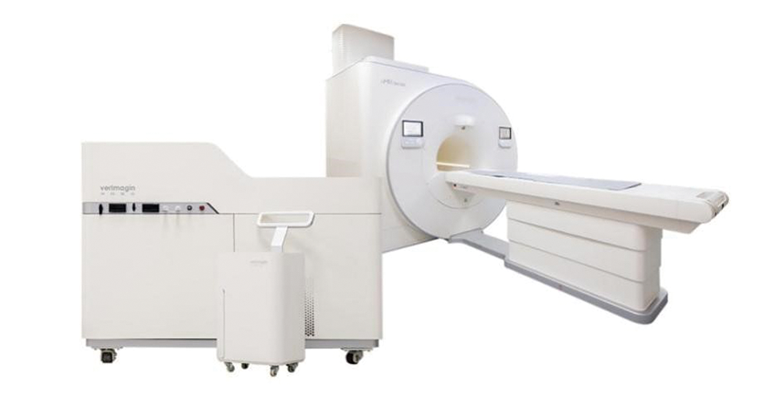
The human lung multi-nuclei magnetic resonance imaging system
According to authoritative statistics, lung cancer has recently ranked first in both incidence and mortality among malignant tumors in China. Enhancing detection technologies for lung diseases, conducting timely screenings, and "tracking down threats within the lungs" are crucial for safeguarding people's health. Traditional clinical imaging devices can reveal obvious tumors and lesions but struggle to detect early changes in gas-blood exchange function and micro-structure within the lungs. In conventional MRI scans, the lungs often appear as an indistinct "black hole," making early diagnosis difficult."If we could develop a more advanced device to 'light up' the lungs, it would greatly enhance detection technology, enabling earlier identification, diagnosis, and treatment of lung diseases and potentially saving millions of lives." Over a decade ago, Xin Zhou, then freshly returned from a research exchange in the United States, envisioned this pioneering equipment. In 2013, as the lead scientist, Zhou spearheaded the launch of the National Major Scientific Instrument and Equipment Development Project in Wuhan, funded by the National Natural Science Foundation, aimed at developing an MRI system for studying major lung diseases. This marked the beginning of a challenging journey.
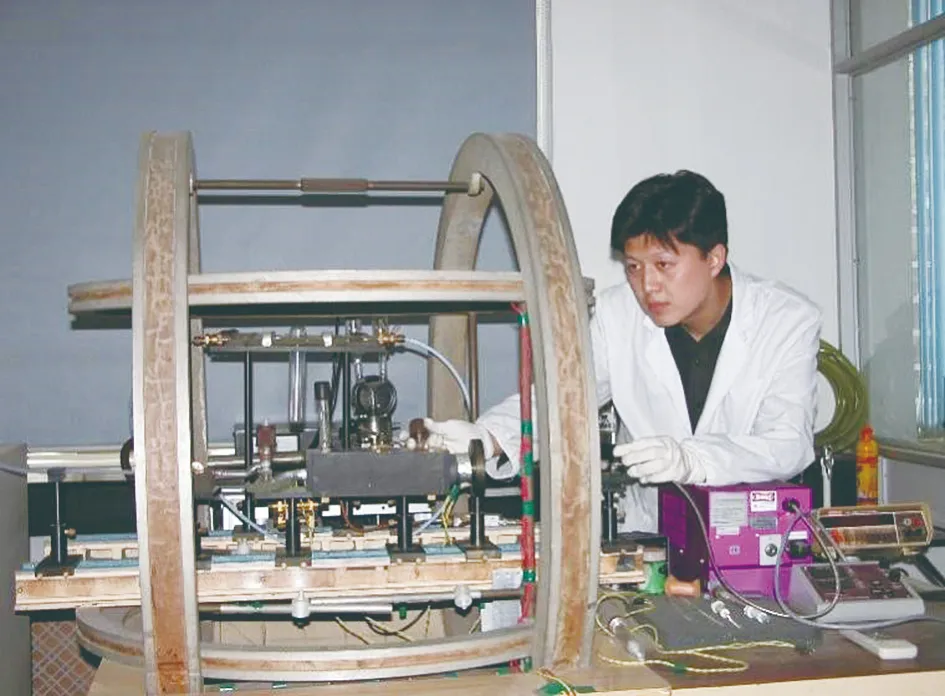
Zhou Xin conducted ultra-sensitive MRI research during his doctoral studies
The key to "tracking down threats" lies in "illuminating" the lung's "black hole." The research team selected two rare, safe, and non-toxic gases helium-3 and xenon-129 with long-lasting MRI signals as potential signal sources. However, they soon noted that helium-3 was costly and insoluble in blood, making it unsuitable for studying gas-blood exchange functions in the lungs. In contrast, xenon-129, with its excellent biological inertness, lipid solubility, and sensitivity to chemical shifts, offered a unique advantage for lung function imaging. Ultimately, the team chose xenon-129 as the lung imaging agent. With broad support, the team made a series of breakthroughs, leading to the development of the "human lung multi-nuclei magnetic resonance imaging system" based on various innovative technologies and equipment. This is the world's first MRI system for gas lung imaging, expanding clinical MRI capabilities from single-nucleus to multi-nucleus imaging. It filled a critical gap in the non-invasive, visual evaluation of lung gas exchange function, pioneering a new field of clinical multi-nuclei MRI imaging in China and placing the nation at the forefront globally. With just one breath of xenon gas, a patient can obtain a 3D MRI image of their lungs within 3.5 seconds. The image clearly shows where the gas has reached in the lungs, providing a detailed view of the lung's micro-structure and gas exchange function. The "human lung multi-nuclei magnetic resonance imaging system," developed by the ultrasensitive magnetic resonance research group of the Innovation Academy for Precision Measurement Science and Technology at the Chinese Academy of Sciences, has effectively solved the scientific challenges of non-destructive, quantitative, and visual detection of lung structure and function, leaving no place for lung disease "killers" to hide. This breakthrough represents a rare original innovation in China's high-end medical equipment field, achieving complete self-sufficiency and control.
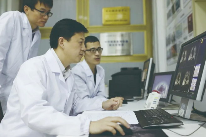
Xin Zhou (second from the left) and his team conducted an experiment
The clinical application of the "human lung multi-nuclei magnetic resonance imaging system" progressed faster than Xin Zhou had anticipated. On January 22, 2020, while Zhou was in Beijing working on medical device registration, he learned that the outbreak in Wuhan was worsening and felt compelled to act. That same evening, he rushed to the airport by taxi from the Chinese Academy of Sciences offices. When the driver heard he was hurrying back to Wuhan to install lung imaging equipment for hospitals, he sped all the way and refused payment. “Returning to Wuhan at a time like this, I can't take your fare,” the driver said, which deeply moved Zhou. Meanwhile, other team members were also converging on Wuhan from various locations, each sensing the battle that lay ahead. As a part of the "national team," the researchers from the Chinese Academy of Sciences knew they could not step back.
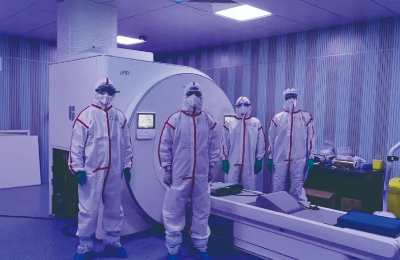
The human lung multi-nuclei magnetic resonance imaging system supported Wuhan's battle against the pandemic
With the support of Dingyu Zhang, the team swiftly installed the "human lung multi-nuclei magnetic resonance imaging system" at Wuhan's Jinyintan Hospital, pioneering the world's first clinical evaluation of lung function in COVID-19 patients. The equipment was also deployed at Tongji Hospital and other frontline hospitals, enabling comprehensive assessments of lung micro-structure and function in over 3,000 COVID-19 patients. Through these studies, they were the first internationally to discover that while discharged patients with mild cases showed no abnormalities in CT imaging or pulmonary function tests, the MRI images from the multi-nuclei system indicated slight damage to ventilation function and significant impairment in gas exchange function. Most of these patients showed progressive improvement in ventilation and gas exchange function by their six-month follow-up. This breakthrough was published in a Science sub-journal, drawing significant attention from international peers. Xin Zhou was also invited by Johns Hopkins University School of Medicine to give an online lecture on the findings. Institutions like the University of Oxford followed up with related studies, highlighting that "gas MRI technology can precisely locate areas of physiological lung impairment."
The impressive performance of China's high-end MRI equipment during the pandemic was no coincidence. From the beginning, Xin Zhou's team was committed to serving public health, focusing on practical applications and driving the system from the lab to real-world use. Through persistent efforts, they developed the "human lung multi-nuclei magnetic resonance imaging system," which became the world's first in its category to obtain a medical device registration certificate and to be applied clinically as a multi-nuclei MRI system capable of gas imaging. The system, developed by the Chinese Academy of Sciences team, has been implemented in over a dozen top-tier hospitals and research institutions across China, including the PLA General Hospital, Shanghai Changzheng Hospital, Wuhan Jinyintan Hospital, and Zhongnan Hospital of Wuhan University. After further optimization, in February 2024, Zhou's team overcame challenges in rapid lung imaging sampling, reducing sampling time to just 3.5 seconds and enhancing image resolution, better catering to lung disease patients who cannot hold their breath for extended periods. This advancement has made the domestically developed "human lung multi-nuclei magnetic resonance imaging system" increasingly accessible and user-friendly.
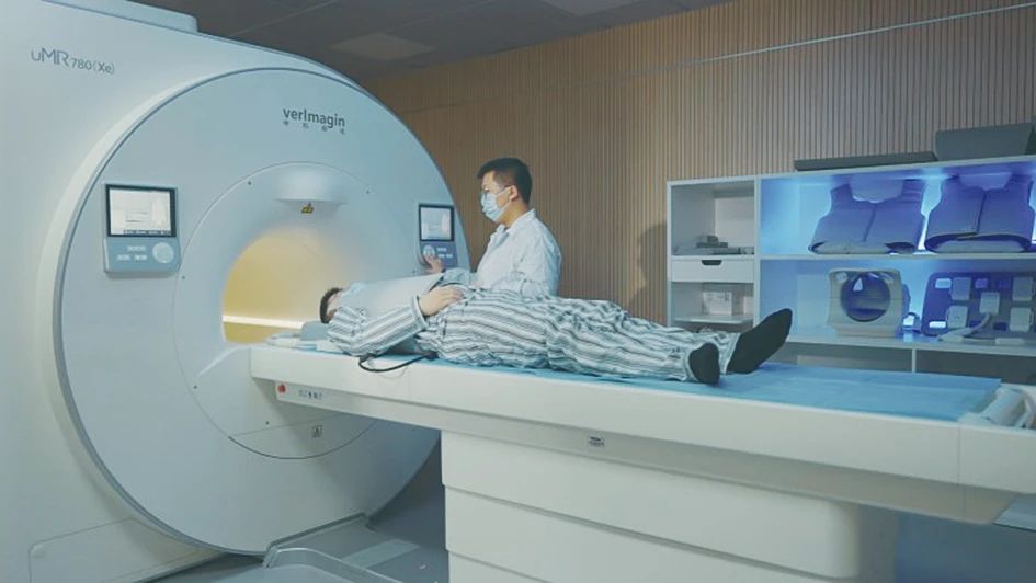
The human lung multi-nuclei magnetic resonance imaging system has entered clinical application
Zhou often recalls a statement by Chaohui Ye, the former director of the Wuhan branch of the Chinese Academy of Sciences, during a graduate lecture: "We must produce high-end domestic medical equipment!" At that time, high-end medical equipment was monopolized by Western multinationals, with exorbitant purchase and maintenance costs, leading to high healthcare expenses for patients. Today, with the widespread adoption of the "human lung multi-nuclei magnetic resonance imaging system," that vision has been realized. In June 2024, the "Multi-nuclei MRI Equipment Development" project was awarded the National Technological Invention Award, Second Class. After the initial excitement, Zhou felt a renewed sense of responsibility. He hopes that the multi-nuclei MRI system, which "lights up" the lungs, will soon become a standard tool in hospitals nationwide—an affordable, accessible, and easily interpretable diagnostic option for all patients, providing a Chinese solution to the challenges of lung disease diagnosis and treatment.
Link: https://news.sciencenet.cn/htmlnews/2024/7/526679.shtm

Innovation Academy for Precision Measurement Science and Technology, CAS.
West No.30 Xiao Hong Shan, Wuhan 430071 China
Tel:+86-27-8719-8631 Fax:+86-27-8719-9291
Email:hanyeqing@wipm.ac.cn
鄂ICP备15017570号-1