
MRI Methods and Techniques for the Lung
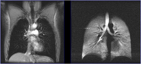
Left image shows conventional proton MR image of lung. The signal originates from the lung's H2O content (about 10^22 hydrogen atoms).
Right image shows a hyperpolarized MR image of lung. The signal originates from helium (3He) gas (about 10^10 atoms).
Lung diseases, especially lung cancer, are a major cause of human death. Thus, imaging techniques that can help identify the onset, type and grade of disease at an early stage is critical. Lungs are normally imaged by x-ray techniques (2D and CT). However, these techniques rely on ionizing radiation, which represents an additional health risk, and do not provide any information that can help assess the function of the lung. The latter is important because an accurate diagnosis should interrogate not only its structure, but whether or not the lung functions (e.g. ventilates) well.
Conventional MRI, based on the use of proton signal, can depict structure and function of brain and other organs, but cannot image lung. This is because the lung consists of:
1) much void space filled with air, which does not yield detectable signal, and
2) its tissue is made of alveolae, which have a very low proton density and short relaxation times due to their porous structure.
In recent years, the technique of hyperpolarized nobles gas MRI has enabled amplifications of the NMR signal by orders of magnitude compared to that of thermal equilibrium. This technique is an excellent tool for the study of lung by MRI and has excellent potential for the diagnosis of lung diseases by measuring associated changes in the lung's structure and function.
Our team develops NMR and MRI techniques based on hyperpolarized noble gases to quantify the changes in lung structure and function associated with various lung diseases. This will, in turn, benefit the medical profession and public health by offering new possibilities for early and accurate diagnosis.

Innovation Academy for Precision Measurement Science and Technology, CAS.
West No.30 Xiao Hong Shan, Wuhan 430071 China
Tel:+86-27-8719-8631 Fax:+86-27-8719-9291
Email:hanyeqing@wipm.ac.cn
鄂ICP备15017570号-1
