


Ultrasensitive Magnetic Resonance Research Group achieve non-invasive assessment of lung function of discharged COVID-19 patients
Recently, Ultrasensitive Magnetic Resonance Research Group, together with Wuhan Tongji Hospital, the Chinese people's Liberation Army General Hospital (301 Hospital), and other units, have applied the self-developed hyperpolarized gas MRI equipment to achieve the non-invasive assessment of lung function injury of discharged COVID-19 patients. This work is published in the international journal Science Advances.
The novel coronavirus pneumonia, which emerged at the end of 2019 (COVID-19), has posed a serious threat to global public health. It has been identified by the WHO (WHO) as a global epidemic. As of November 19, 2020, novel coronavirus pneumonia cases in more than 200 countries and regions had more than 55 million 920 thousand confirmed cases, and the cumulative number of deaths was over 1 million 340 thousand. It was found that some patients still experienced different degrees of fatigue, dyspnea and other symptoms after discharge.
Lung gas MRI technology overcomes the technical challenge of clinical proton (1H) MRI on the "blind area" of lung cavity, and provides a new medical equipment for quantitative and visual detection of major lung diseases. The equipment can be used for quantitative and visual evaluation of lung function of patients with COVID-19, effectively solve the problem that conventional clinical detection equipment (CT, chest X-ray) has ionizing radiation and cannot realize quantitative detection of lung function, and breaks through the bottleneck of high-performance imaging instrument.
Ultrasensitive Magnetic Resonance Research Group applied self-developed gas MRI equipment to achieve the quantitative and visual assessment of lung microstructure, ventilation and gas-blood exchange function of discharged COVID-19 patients for the first time in the world. It was found that although the lung CT images and the parameters of discharged patients’ lung function with common diseases were not abnormal, their gas MRI images showed slight impairment of ventilation function and obvious impairment of gas-blood exchange function. The ventilation and gas-blood exchange functions of most discharged patients with common diseases showed progressive improvement at the sixth month of follow-up.
This research not only provides scientific and technological support for the clinical rehabilitation of discharged patients with new coronary pneumonia, but also is an important supplement to existing clinical imaging technology.
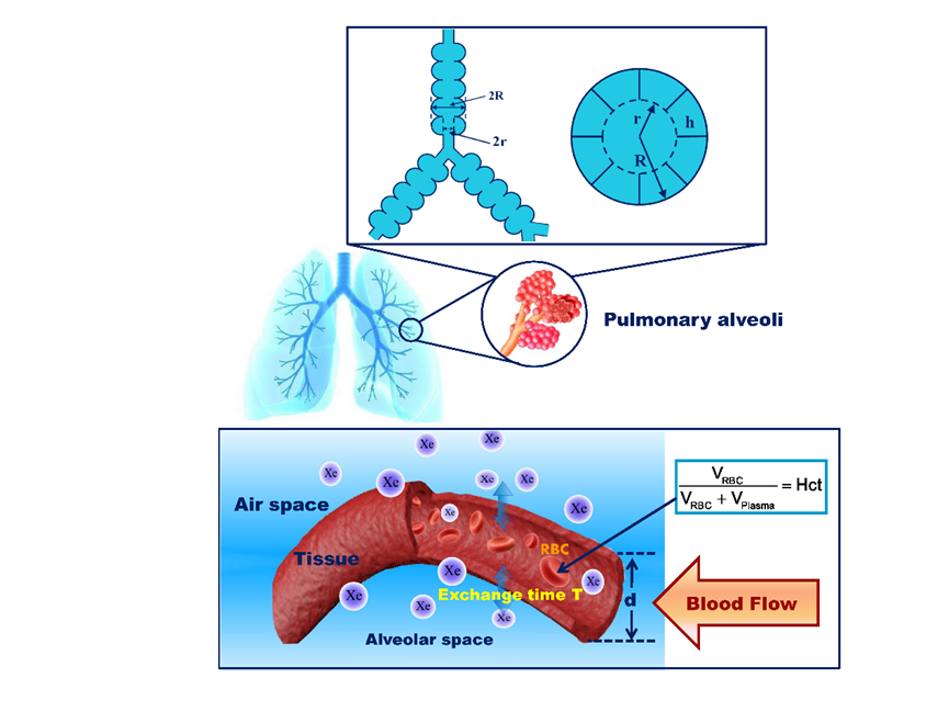
Diagram of lung microstructure and gas-blood exchange quantitative inspection based on hyperpolarized 129Xe gas MRI technology.
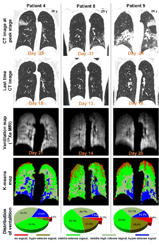
Typical lung images of three COVID-19 patients discharged from hospital.
From top to bottom: CT images of the lungs in the acute phase of pneumonia, CT images of the lungs at the end of the recovery period (before and after discharge), corresponding lung gas MRI images and clustered lung ventilation function images.

Innovation Academy for Precision Measurement Science and Technology, CAS.
West No.30 Xiao Hong Shan, Wuhan 430071 China
Tel:+86-27-8719-8631 Fax:+86-27-8719-9291
Email:hanyeqing@wipm.ac.cn
鄂ICP备15017570号-1
